Anti-AAK1 Polyclonal Antibody
Ordering
Available Variants| Availability & Stock | Pack | SKU | Price (EUR) |
|---|---|---|---|
|
Available – Dispatch in 14 days
Global Network - International delivery
|
20ul | CAB.12987-20ul | €100.00 |
|
Available – Dispatch in 14 days
Global Network - International delivery
|
50ul | CAB.12987-50ul | €500.00 |
|
Available – Dispatch in 14 days
Global Network - International delivery
|
100ul | CAB.12987-100ul | €500.00 |
|
Available – Dispatch in 14 days
Global Network - International delivery
|
100ul(PBS Only) | CAB.12987-100ul(PBS Only) | €500.00 |
| Catalog No | CAB.12987 |
|---|---|
| Product Name | Anti-AAK1 Polyclonal Antibody |
| Isotype | IgG |
| Calculated MW | 94/104kDa |
| Immunogen | Recombinant protein of human AAK1 |
| Public Immunogen Range | 1-350aa |
| Host | Rabbit |
| Clone Type | Polyclonal Antibody |
| Reactivity | Cow(99.1%);Pig(99.1%) |
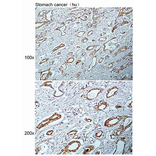
| Application | IHC-P |
| Subcellular Location | Cytoplasm Nucleus |
| Purification Method | CABfinity purification |
| Storage Buffer | PBS with 50% Glycerol,0.03% Proclin300,0.5% BSA,pH 7.3. |
| Storage | Store at -20℃. Avoid freeze / thaw cycles. |
Background
Regulates clathrin-mediated endocytosis by phosphorylating the AP2M1/mu2 subunit of the adaptor protein complex 2 (AP-2) which ensures high CABfinity binding of AP-2 to cargo membrane proteins during the initial stages of endocytosis (PubMed:17494869, PubMed:11877457, PubMed:11877461, PubMed:12952931, PubMed:14617351, PubMed:25653444).
Protocol / Instructions
The Anti-AAK1 Polyclonal Antibody is a rabbit-derived antibody that recognizes the AAK1 protein in cow and pig species. This antibody has been validated for use in Immunohistochemistry-Paraffin (IHC-P) applications.
Reagents or buffers: Xylene, ethanol, antigen retrieval buffer, blocking buffer, primary antibody (Anti-AAK1 Polyclonal Antibody), secondary antibody, DAB substrate.
Sample preparation: Paraffin-embedded tissue sections are deparaffinized in xylene and rehydrated through a series of ethanol washes. Antigen retrieval is performed using a suitable buffer.
Antibody incubation: The primary antibody is applied at a recommended dilution of 1:50-200 and incubated for 1 hour at room temperature or overnight at 4°C.
Washing or detection: The sections are washed with a suitable buffer, and a secondary antibody conjugated to a detection enzyme (e.g., HRP) is applied. The signal is visualized using a DAB substrate.
Technical notes: Optimize the primary antibody dilution and incubation time to achieve the best signal-to-noise ratio. Use a suitable negative control to verify the specificity of the staining.
Precautions
Store at -20°C. Handle with care, and avoid repeated freeze-thaw cycles. Optimize antibody dilution and incubation conditions for best results.
Validation Images