Anti-AAAS Polyclonal Antibody
Ordering
Available Variants| Availability & Stock | Pack | SKU | Price (EUR) |
|---|---|---|---|
|
Available – Dispatch in 14 days
Global Network - International delivery
|
50ul | CAB.13682-50ul | €200.00 |
|
Available – Dispatch in 14 days
Global Network - International delivery
|
100ul | CAB.13682-100ul | €335.00 |
| Catalog No | CAB.13682 |
|---|---|
| Product Name | Anti-AAAS Polyclonal Antibody |
| Isotype | IgG |
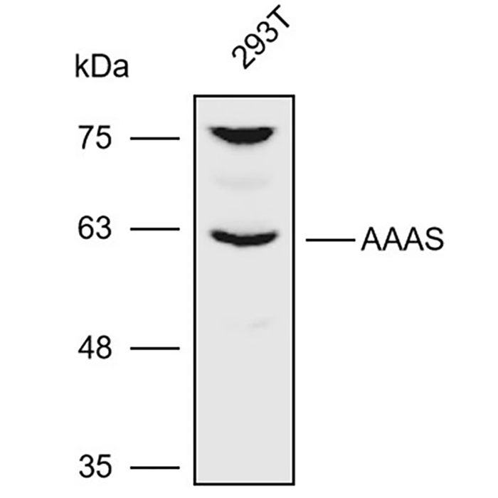
| Calculated MW | 60kDa |
| Immunogen | Recombinant protein of human AAAS |
| Public Immunogen Range | N/A |
| Host | Rabbit |
| Clone Type | Polyclonal Antibody |
| Reactivity | Mouse;Human |
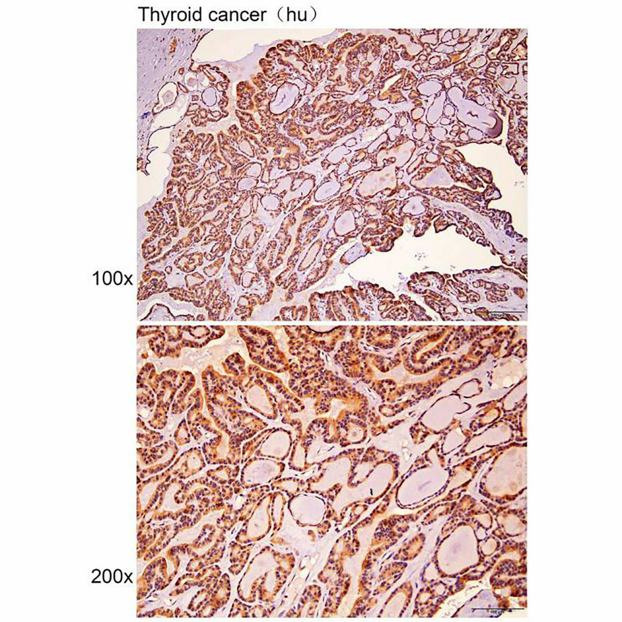
| Application | IHC-P;WB |
| Subcellular Location | Cytoplasm |
| Purification Method | CABfinity purification |
| Storage Buffer | pH7.4 PBS,0.05% NaN3,40% Glycerol. |
| Storage | Store at -20℃. Avoid freeze / thaw cycles. |
Background
The protein encoded by this gene is a member of the WD-repeat family of regulatory proteins and may be involved in normal development of the peripheral and central nervous system. The encoded protein is part of the nuclear pore complex and is anchored there by NDC1. Defects in this gene are a cause of achalasia-addisonianism-alacrima syndrome (AAAS), also called triple-A syndrome or Allgrove syndrome. Two transcript variants encoding different isoforms have been found for this gene.
Protocol / Instructions
The Anti-AAAS Polyclonal Antibody is a rabbit-derived antibody that recognizes the AAAS protein in human and mouse samples. This antibody has been validated for use in Western Blotting (WB) and Immunohistochemistry-Paraffin (IHC-P) applications.
For Western Blotting, the recommended dilution is 1:200-1000. The following reagents and buffers are required: sample buffer, running buffer, transfer buffer, and blocking buffer. Sample preparation involves lysing cells or tissues in sample buffer, followed by separation on an SDS-PAGE gel and transfer to a membrane. The membrane is then blocked with blocking buffer and incubated with the Anti-AAAS Polyclonal Antibody. CABter washing, the membrane is incubated with a secondary antibody conjugated to a detection system. Technical notes: the optimal dilution and incubation time may vary depending on the specific sample and experimental conditions.
For Immunohistochemistry-Paraffin, the recommended dilution is 1:10-100. The following reagents and buffers are required: xylene, ethanol, antigen retrieval buffer, and blocking buffer. Sample preparation involves deparaffinizing and rehydrating tissue sections, followed by antigen retrieval and blocking with blocking buffer. The sections are then incubated with the Anti-AAAS Polyclonal Antibody, followed by washing and incubation with a secondary antibody conjugated to a detection system. Technical notes: the optimal dilution and incubation time may vary depending on the specific tissue and experimental conditions.
Precautions
Store at -20°C, handle with care, use appropriate controls, and optimize antibody dilution and incubation time for best results.
Validation Images