Anti-A4GALT Polyclonal Antibody
Ordering
Available Variants| Availability & Stock | Pack | SKU | Price (EUR) |
|---|---|---|---|
|
Available – Dispatch in 14 days
Global Network - International delivery
|
50ul | CAB.25448-50ul | €200.00 |
|
Available – Dispatch in 14 days
Global Network - International delivery
|
100ul | CAB.25448-100ul | €335.00 |
| Catalog No | CAB.25448 |
|---|---|
| Product Name | Anti-A4GALT Polyclonal Antibody |
| Isotype | IgG |
| Calculated MW | 40kDa |
| Immunogen | Recombinant protein of human A4GALT |
| Public Immunogen Range | N/A |
| Host | Rabbit |
| Clone Type | Polyclonal Antibody |
| Reactivity | Mouse;Rat;Human |
| Application | WB |
| Subcellular Location | Golgi apparatus |
| Purification Method | CABfinity purification |
| Storage Buffer | pH7.4 PBS,0.05% NaN3,40% Glycerol. |
| Storage | Store at -20℃. Avoid freeze / thaw cycles. |
Background
The protein encoded by this gene catalyzes the transfer of galactose to lactosylceramide to form globotriaosylceramide, which has been identified as the P(k) antigen of the P blood group system. This protein, a type II membrane protein found in the Golgi, is also required for the synthesis of the bacterial verotoxins receptor. Alternatively spliced transcript variants have been found for this gene.
Protocol / Instructions
The Anti-A4GALT Polyclonal Antibody is a rabbit-derived antibody that recognizes the A4GALT protein in mouse, rat, and human samples. This antibody has been validated for use in Western Blotting (WB) applications.
For Western Blotting, the recommended dilution of the Anti-A4GALT Polyclonal Antibody is 1:200-1000.
Reagents or buffers required include a suitable lysis buffer for sample preparation, a transfer buffer for membrane transfer, and a blocking buffer to reduce non-specific binding.
Sample preparation involves lysing cells or tissues in a lysis buffer, followed by protein quantification and separation by SDS-PAGE.
The separated proteins are then transferred to a membrane, which is blocked and incubated with the Anti-A4GALT Polyclonal Antibody.
CABter washing to remove unbound antibody, the membrane is incubated with a secondary antibody conjugated to a detection system, such as HRP or a fluorescent dye.
Technical notes: Optimize the antibody dilution and incubation time to achieve the best signal-to-noise ratio. The use of a positive control, such as a recombinant A4GALT protein, can help verify the specificity of the antibody.
Precautions
Store at -20°C, handle with gloves, and optimize antibody dilution for best results.
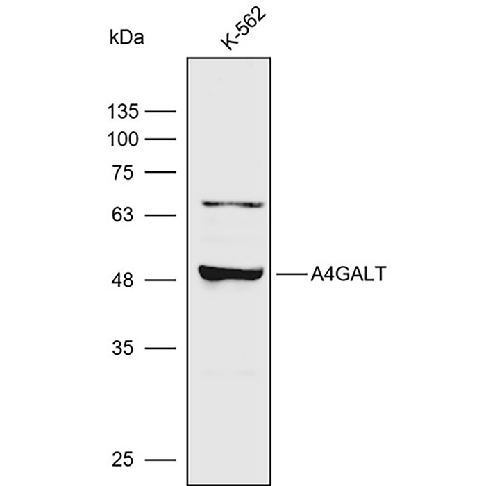
Validation Images